Rib Cage Muscles Anatomy / Labeled Lungs Diagram With Rib Cage Rib Cage Anatomy ... : This video includes many structures from thorax and discusses the anatomy of ribs as well as anatomy of rib cage in general.
Rib Cage Muscles Anatomy / Labeled Lungs Diagram With Rib Cage Rib Cage Anatomy ... : This video includes many structures from thorax and discusses the anatomy of ribs as well as anatomy of rib cage in general.. Structure of a typical rib: Measuring rib cage and abdominal movement is the most common technique for assessing respiratory effort in laboratory sleep studies. This is where the gto comes into play. Chest bone rib cage landmark diagram. Anterior view of the lungs and ribcage in a transparent female torso stock illustration these pictures of this page are about:human anatomy rib cage muscles.
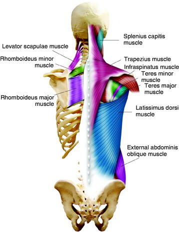
Your ribs form a protective cage that encloses many of your delicate internal organs, such as your heart and lungs. They are extremely light, but highly resilient; Another shoulder positioning muscle that can be observed on. Various skeletal muscles are attached to the rib cage. Structure of a typical rib:
Posterior and Posterolateral Access to the Thoracic Spine ... from neupsykey.com Skeletal muscles attached to the rib cage: Struggling with learning muscle attachments? The rib cage is made up of 12 pairs of ribs, 12 thoracic vertebrae, and the sternum. Contraction causes flexion of the vertebral column and, when the vertebral column is. We hope you will use this picture in the study and helping your research. They are more involved in forced expiration and coughing to forcibly shrink the chest and. The rib cage, shaped in a mild cone shape and more flexible than most bone sets, is made up of varying elements such as the thoracic vertebra, 12 equally paired ribs, costal cartilage, and held together anteriorly by the sternum. On a muscular person when the muscles stretch, we see some of the lower ribs in the front and also in the back.
Anatomy is the amazing science.
Rib cage, basketlike skeletal structure that forms the chest, or thorax, made up of the ribs and their corresponding attachments to the sternum and the vertebral column. Various skeletal muscles are attached to the rib cage. The rib cage, shaped in a mild cone shape and more flexible than most bone sets, is made up of varying elements such as the thoracic vertebra, 12 equally paired ribs, costal cartilage, and held together anteriorly by the sternum. In vertebrate anatomy, ribs (latin The rib cage is often simplified as an oval shape. Muscle spasms located in the rib cage are often observed in people who strain or overwork their upper body muscles. Structure of a typical rib: Anterior view of the lungs and ribcage in a transparent female torso stock illustration these pictures of this page are about:human anatomy rib cage muscles. 1887 human anatomy print of the rib cage and sternum. The rib cage is a primarily protective structure, encircling the heart and lungs. The thorax is anatomical structure supported by a skeletal framework (thoracic cage) and thoracic cage is formed by bones and cartilage osseocartilaginous. It can help you understand our world more detailed and specific. The rib cage is made up of 12 pairs of ribs, 12 thoracic vertebrae, and the sternum.
Some extend from above and draw the. The thorax is anatomical structure supported by a skeletal framework (thoracic cage) and thoracic cage is formed by bones and cartilage osseocartilaginous. For example, flexor, extensor, adductor and abductor are names associated with the action of the muscle. But for an anatomy study, it's not. Another shoulder positioning muscle that can be observed on.
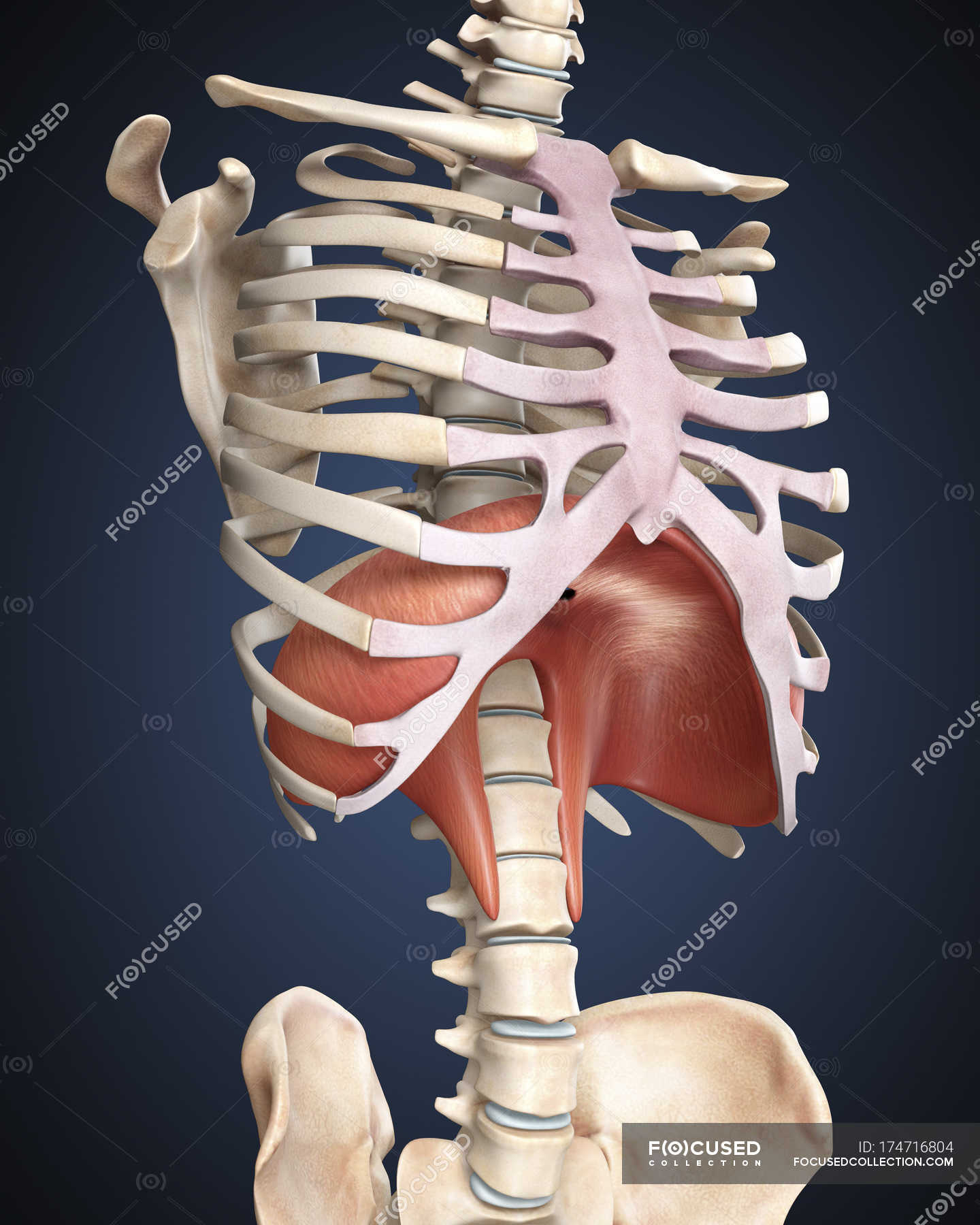
Medical illustration of human diaphragm in rib cage ... from st.focusedcollection.com Rib somatic dysfunction rib dysfunctions are grouped into two categories: The rib cage is often simplified as an oval shape. The rib cage, shaped in a mild cone shape and more flexible than most bone sets, is made up of varying elements such as the thoracic vertebra, 12 equally paired ribs, costal cartilage, and held together anteriorly by the sternum. They articulate with the vertebral column posteriorly, and terminate they also have a role in ventilation; Anatomy is the amazing science. Another shoulder positioning muscle that can be observed on. Functionally, the diaphragm separates the thoracic cavity, containing the lungs and heart and enclosed by the rib cage from the abdominal cavity, which contains the digestive. So what parts of the rib cage show up on the surface?
Your rib cage plays an important role in respiration, expanding and contracting as your respiratory muscles, including your diaphragm, work to help you breathe.
Check out our muscle anatomy reference charts to learn faster! Structure of a typical rib: Contraction causes flexion of the vertebral column and, when the vertebral column is. The ribcage is made to be flexible and springy so the lungs can fill and deflate easily. So what parts of the rib cage show up on the surface? The rib cage is often simplified as an oval shape. On a muscular person when the muscles stretch, we see some of the lower ribs in the front and also in the back. They are more involved in forced expiration and coughing to forcibly shrink the chest and. The rib cage is a primarily protective structure, encircling the heart and lungs. Rib 2 is thinner and longer than rib 1 and has two articular facets on the head as normal. Create your own flashcards or choose from millions created our most recent study sets focusing on rib cage muscles will help you get ahead by allowing you to study whenever and wherever you want. They articulate with the vertebral column posteriorly, and terminate they also have a role in ventilation; The rib cage is the arrangement of ribs attached to the vertebral column and sternum in the thorax of most vertebrates, that encloses and protects the vital organs such as the heart, lungs and great vessels.
This is where the gto comes into play. For example, flexor, extensor, adductor and abductor are names associated with the action of the muscle. Skeletal muscles attached to the rib cage: The rib cage surrounds the lungs and the heart, serving as an important means of bony protection for these vital organs. Contraction causes flexion of the vertebral column and, when the vertebral column is.
Labeled Lungs Diagram With Rib Cage Rib Cage Anatomy ... from i.pinimg.com While muscle spasms may occur over the entire body, muscle spasms under the rib cage may be cause for concern as they might be an indication of serious medical conditions. Check out our muscle anatomy reference charts to learn faster! The rib cage is often simplified as an oval shape. Skeletal muscles attached to the rib cage: Rib cage, basketlike skeletal structure that forms the chest, or thorax, made up of the ribs and their corresponding attachments to the sternum and the vertebral column. Muscles of the thoracic wall contain those that fill and support the intercostal spaces, those that pass between the sternum and the ribs, and those that cross several ribs between costal attachments. The ribcage is made to be flexible and springy so the lungs can fill and deflate easily. Some extend from above and draw the.
Your ribs form a protective cage that encloses many of your delicate internal organs, such as your heart and lungs.
Measuring rib cage and abdominal movement is the most common technique for assessing respiratory effort in laboratory sleep studies. Skeletal muscles attached to the rib cage: Ribs are not merely armour for the organs inside our torsos, as we rib fractures are a common and very painful injury, with the middle ribs the most likely ones to get the muscles that move the ribcage itself are the intercostal muscles. They are each attached to the ribs. The ribcage is made to be flexible and springy so the lungs can fill and deflate easily. Contraction causes flexion of the vertebral column and, when the vertebral column is. Various skeletal muscles are attached to the rib cage. Your ribs form a protective cage that encloses many of your delicate internal organs, such as your heart and lungs. Contributing to their role in protecting the internal thoracic organs. Musculoskeletal considerations of the rib cage. Muscle spasms located in the rib cage are often observed in people who strain or overwork their upper body muscles. • raise rib cage for inhaling & depresses rib cage for exhaling. We hope you will use this picture in the study and helping your research.
It has clear front side and back planes rib cage muscles. Muscle spasms located in the rib cage are often observed in people who strain or overwork their upper body muscles.


0 Komentar